

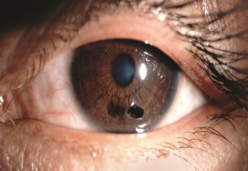
As pictured above, a corneal foreign body is a foreign object that adheres to, or embeds into, the tissue of the cornea, the clear dome at the front of the eye. Symptoms include sudden eye pain, decreased visual acuity, light sensitivity, and redness of the white of the eye. Most corneal foreign bodies can be successfully removed at the slit lamp in the office with the use of topical anesthetic drops and forceps or another corneal foreign body removal tool. Prompt removal of any foreign body minimizes the risk of infection, inflammation, scarring, and permanent vision loss. After removal of the offending object, patients will almost always receive topical ophthalmic antibiotic drops and/or ointments to prevent infection from whatever microbes might have existed on the foreign body removed from the surface of the cornea.
Following the removal of large, or deeply embedded corneal foreign bodies, a therapeutic bandage contact lens and sometimes an amniotic membrane, may be placed on the cornea to reduce pain and discomfort, diminish scarring, and protect the cornea during healing. If you suspect you have a corneal foreign body, call 314-860-2020.
The corneal foreign body depicted below is a large flat foreign body that has only been in the eye for a short time as there is very little swelling around the foreign body and very little redness in the eye. This is likely vegatative matter and these are typically straightforward to remove as long as the patient is seen promptly for removal. These patients are sometimes seen months or years later with a recurrent corneal erosion, as this type of foreign matter has a tendency to cause the skin of the cornea to become less adherent to the underlying stromal layer of the cornea.
RECURRENT CORNEAL EROSION
The image below displays an area of the cornea that has surface skin that is essentially peeling off. This could be occurring because of a history of physical truama, a chemical trauma, a paper cut abrasion, a fingernail abrasion, or vegetative matter that caused the corneal skin to no longer be as adherent to the underlying membrane that used to hold the skin tightly to the surface of the eye. This can often benefit from the use of an amniotic membrane as discussed earlier. Sometimes, however, more invasive procedures become necessary like laser phototherapeutic keratectomy, or PTK.

METALLIC CORNEAL FOREIGN BODIES
The image below depicts an embedded metallic corneal foreign body below this patient's visual axis. The surrounding corneal swelling and the obvious rust in this image support the fact that this foreign body has been ignored for too long. Because of this delay, this now will require removal of the actual foreign body as well as removal of the rust ring that will likely remain in the corneal tissue after the foreign body has been removed. If you, or a family member, have been grinding metal, working around metal shavings, or working removing rust from a metal surface, and you believe you may have a metallic foreign body in either eye, please call Dr. Sullivan or Dr. Wilkins at 314-860-2020 to have the foreign body removed as quickly as possible.
